Last reviewed October 29, 2026 · Dr. Ilseop Ahn (board-certified periodontist, specialist in integrated dentistry)
At a glance
Good for
Where a tooth is missing or cannot be saved
Treatment flow
Check first whether the tooth can be saved → 3D CT measurement → guided placement → crown → regular check-ups
Duration and limits
Usually a few months until the crown. Check-ups at 1, 3, and 6 months after the crown, then every 6 months
Details
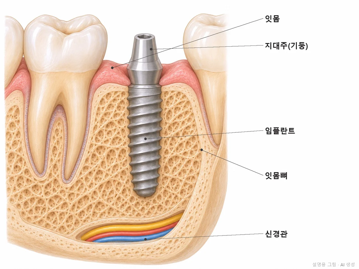
A dental implant places an artificial root where a tooth is missing or cannot be saved, and a tooth is then placed on top. If your current tooth can be saved, you do not have to get one. Before any extraction, Ceramic Dental Clinic measures gum depth, tooth mobility, and bone height to check first whether the tooth can be saved. If an implant is needed, a board-certified periodontist, who specializes in gums and bone, measures the bone to the millimeter with 3D CT and places the implant with a navigation guide, and the same dentist also handles the crown and later check-ups. From placement to the crown usually takes a few months, depending on the bone. After the crown, check-ups are at 1, 3, and 6 months, then every 6 months (every 3 months for higher-risk patients).
01°
Even with the same 'implant consultation,' the conclusion differed from person to person
Teeth that could be saved were kept, sites with too little bone received bone grafts during implant placement, and for a patient who had lost most teeth, we first selected which remaining teeth to keep. Here are three typical situations from real cases.
CASE01
A molar so loose the patient wondered whether it had to come out
Gum treatment instead of an implant · Gum depth measured again before and after treatment to confirm the tooth could be kept
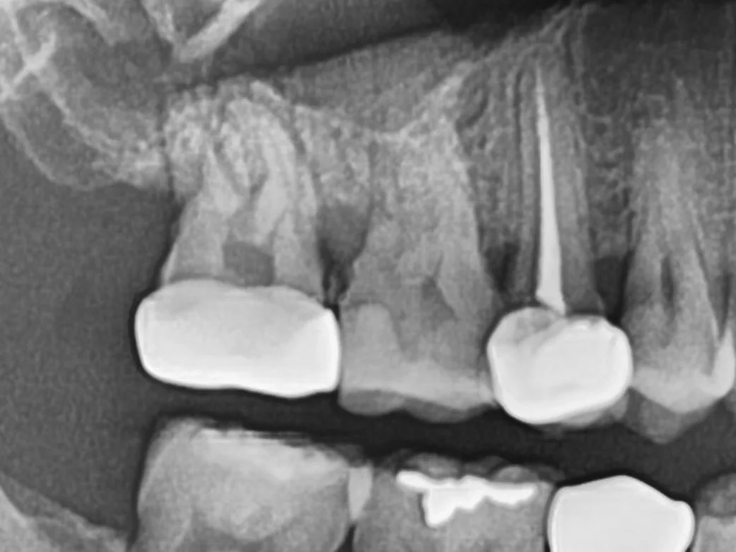
Before · Jun 2020
Before treatment, panoramic X-ray. The gum bone had dropped behind the crowned upper right second molar.
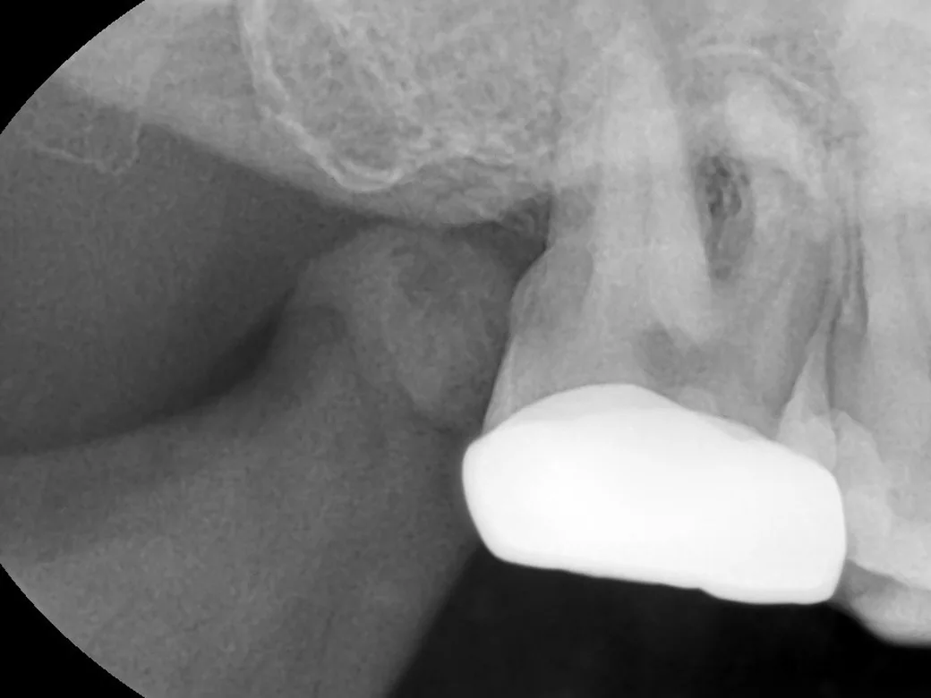
Day of gum treatment · Oct 2020
Periapical X-ray on the day of gum treatment. The patient came in with pain because the bone behind the tooth had dropped, and on the same day we did gum treatment to scrape tartar and bacterial film off the root surface. When we checked 10 days later, the mobility had decreased.
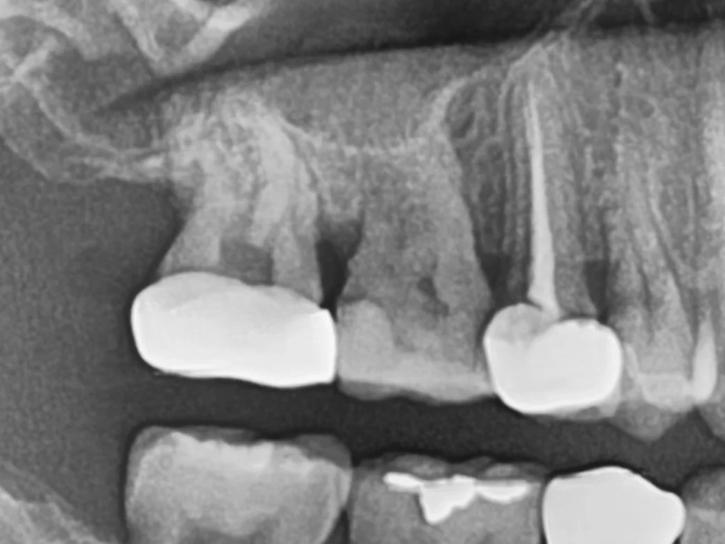
3 years 3 months later · Jan 2024
Panoramic X-ray 3 years 3 months later. The molar that was not extracted is still in use, and when matched to the same scale as the first image, the bone had not dropped noticeably further. Because this is a panoramic X-ray, precise millimeter comparison is difficult.
Condition
A molar that was loose because gum disease had lowered the bone
Chosen
Removal of tartar and bacterial film below the gums, re-evaluation, and regular care
Not done
Extraction and implant (re-evaluation showed the tooth could be kept)
Upper molar site with slightly too little bone because of the space above (the maxillary sinus)
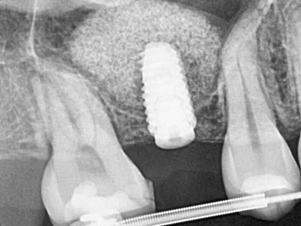
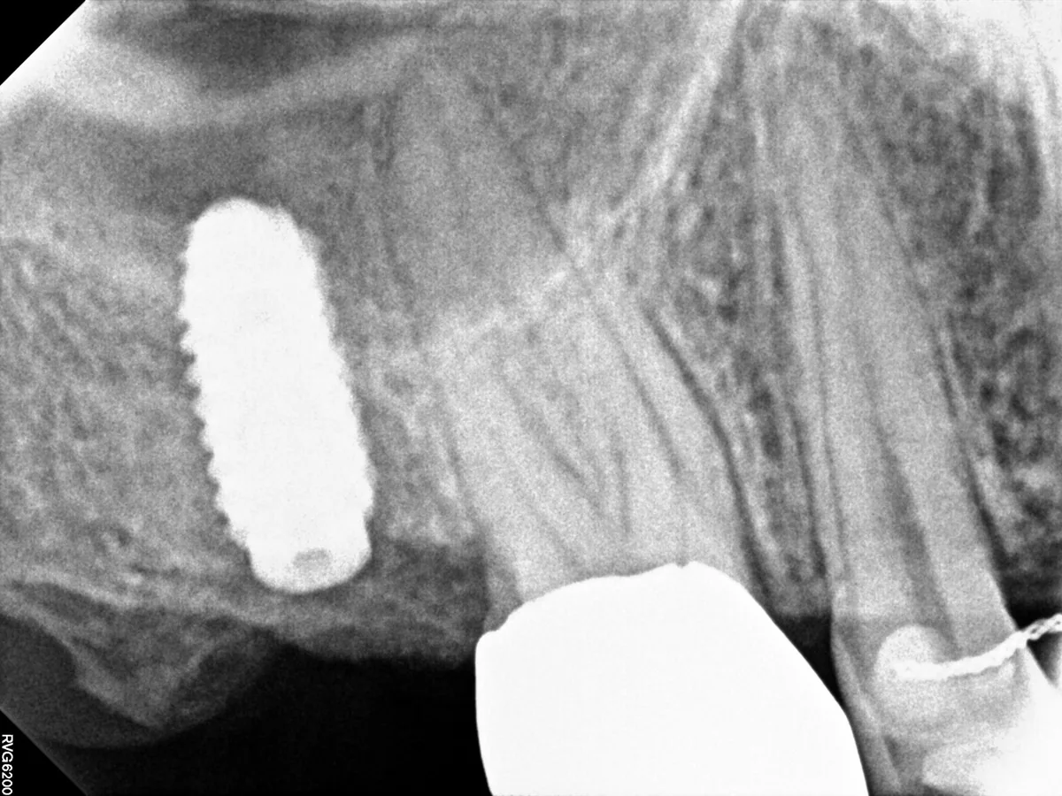
Sinus floor gently lifted through the implant hole, with placement on the same day
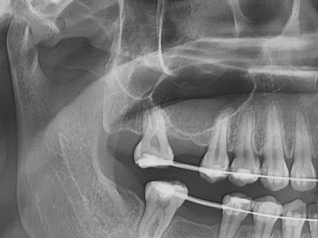
Before · Aug 2023
Before treatment, X-ray (panoramic). Because of the maxillary sinus, the bone at the upper right first molar site was low and slightly short of the implant length. There was enough to hold the implant when placed.
13 days after placement · Apr 2024
X-ray 13 days after placement. The sinus floor was gently lifted through the implant hole and graft material was added, forming a round dome above the implant tip.
5 and a half months later · Oct 2024
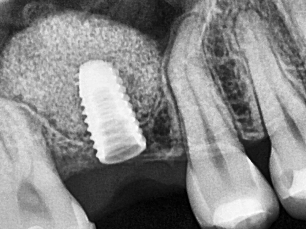
X-ray 5 and a half months later. We confirmed the dome-shaped graft area had remained and took the impression for the crown.
Condition
Upper molar, about 3 mm of bone height remaining
Chosen
Sinus floor lifted through the implant hole, placement on the same day
Not done
Major surgery opening a window from the side (only a small lift was needed)
An upper jaw where most remaining teeth were loose
Chose the teeth to keep first, lived with temporary teeth, and rebuilt the whole arch
Before · Jul 2020
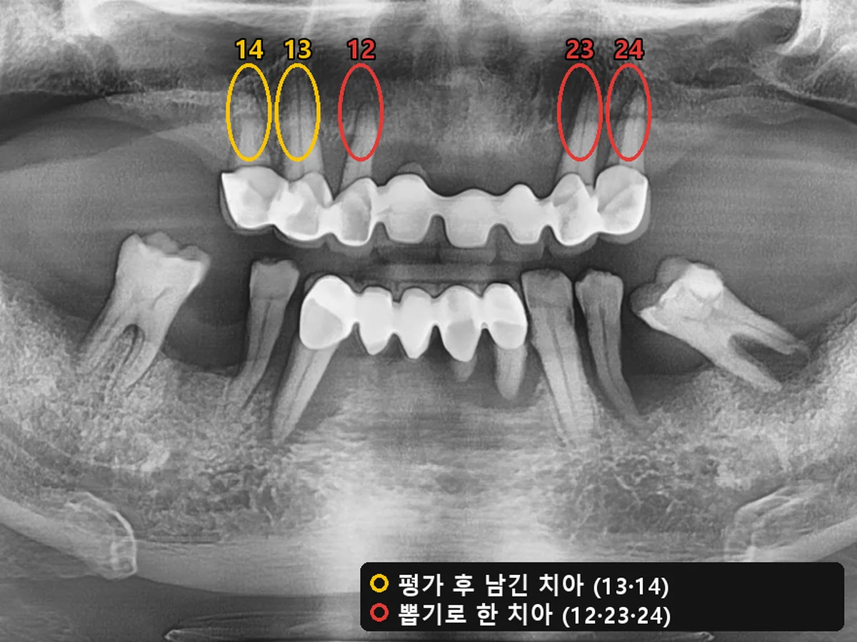
Before treatment, panoramic X-ray and map of teeth to keep. After assessing each tooth, we decided to keep #13 and #14 and extract #12, #23, and #24.
Check-up · Jun 2022
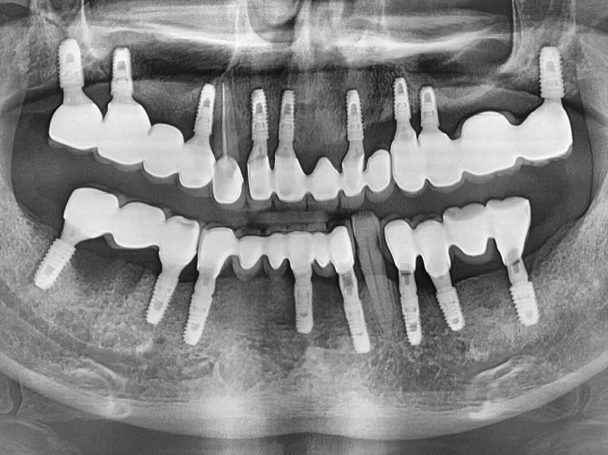
Panoramic X-ray taken at a check-up after the final restorations. The whole upper jaw was rebuilt with the kept teeth and implants, and the biting height was adjusted. The lower jaw was treated as well.
Condition
Advanced gum disease, with most upper teeth loose
Chosen
2 teeth kept · 9 implants in the upper jaw · lived with temporary teeth
Duration
About 9 months
Note
Cleaning under the restoration is difficult, so 3-month check-ups are essential
* Photos were taken under the same conditions and published with the patients' consent. Treatment results and side effects may vary from person to person.
02°
Depending on your concern, see this page first
Implant and gum care decisions differ from case to case. We wrote more detail on the page that matches your concern.
A specialist in gums and bone places the implant and also handles the crown and check-ups
Periodontics is the specialty that deals with the gums, gum bone, bone grafts, and tissue around implants, and integrated dentistry is the specialty that looks after the whole tooth, from cavities and root canal treatment to crowns. I hold both qualifications, so I follow your care through from building bone to the crown placed on top and the care afterward.
BasisBoard-certified periodontist · Specialist in integrated dentistry (Ministry of Health and Welfare) · See the original certificates
Key Point. 2
If a tooth can be saved, we save it
Before any extraction, we measure gum depth, mobility, and bone height, show them to you on screen, and explain both the option of saving the tooth and the option of an implant. In many cases, it is not too late to measure again after gum treatment and then decide.
BasisGum depth measured for each tooth · Digital panoramic X-ray · 3D CT
Key Point. 3
We plan to the millimeter with CT and place implants as planned with a guide
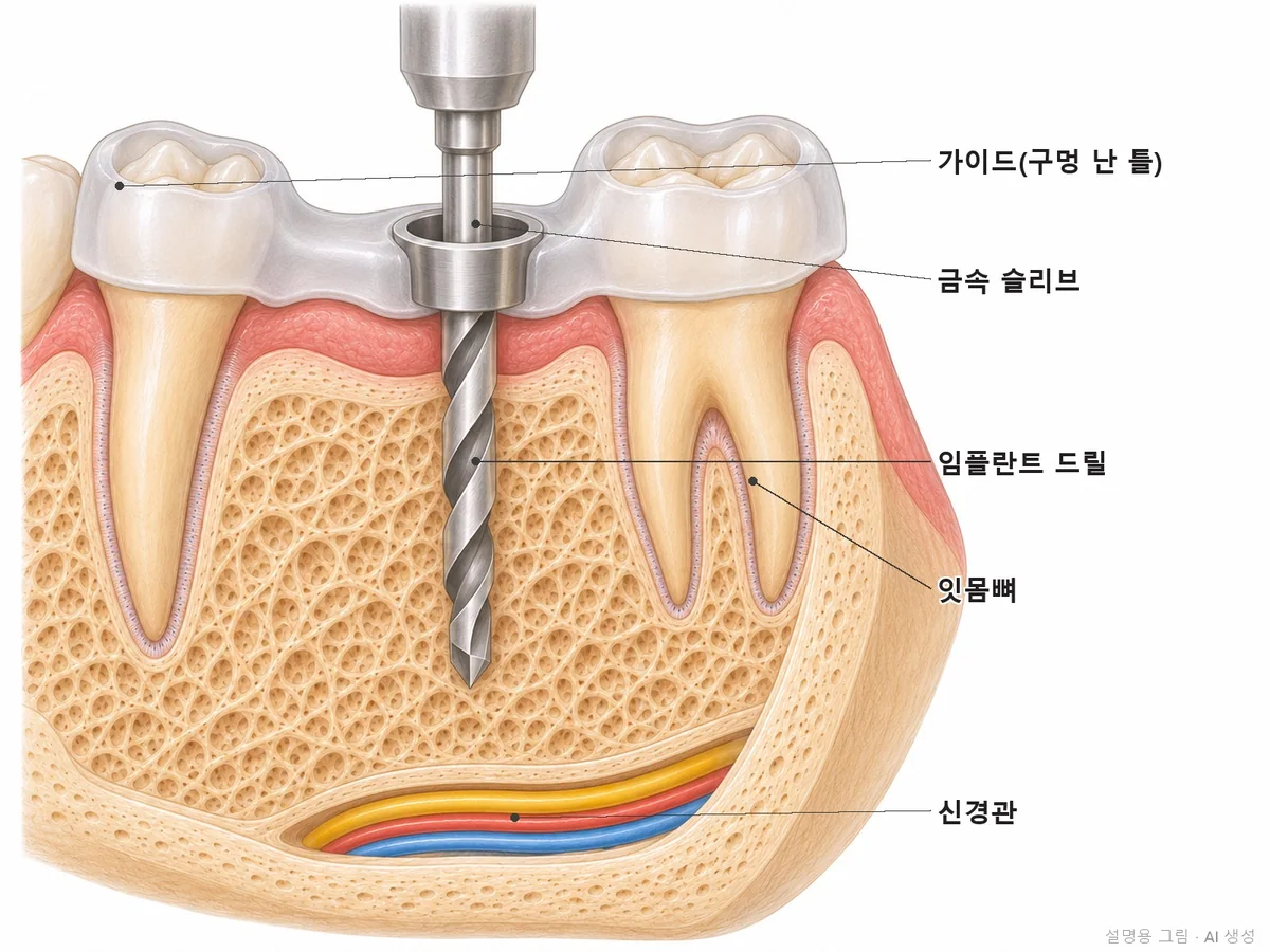
With 3D CT we measure bone height and thickness and the distance to the lower jaw nerve and the maxillary sinus, and decide the implant position, angle, and depth in advance. To place it as planned, we use a navigation guide (surgical template).
Check-ups are at 1, 3, and 6 months after the crown, then every 6 months. People who have had gum disease, smoke, have diabetes, or have several implants are seen every 3 months.
We disclose the products we use and the insurance criteria
We use Osstem implants, and we have also put together a separate summary of who is eligible for implants under Korea's National Health Insurance at age 65 or older and how much patients pay.
Save the tooth or get an implant? We decide by looking at these numbers
We do not decide by a single number. We look at the five factors below together and explain how things are likely to go if the tooth is saved versus extracted.
Gum depth · A healthy gum pocket is usually 3 mm or less. If many sites are over 5 mm and bleed, gum treatment is needed first.
Mobility · We see how much, and in which direction, the tooth moves when pushed by hand. If it moves even when pressed up and down, it is often hard to keep.
Remaining bone · With panoramic X-rays and CT, we see how much of the root length is still surrounded by gum bone.
Root and tooth condition · If the root is cracked or decay has gone deep below the gums, the tooth is hard to use for long even if saved.
Overall plan · We also consider whether saving this one tooth helps the neighboring teeth and future restoration plans.
Teeth loose from gum disease sometimes become less loose when measured again after gum treatment, so it is not too late to decide then.
05°
How implant treatment works
There are seven steps, from exam to check-ups. For each step, we explain what we look at and why, with real clinical photos.
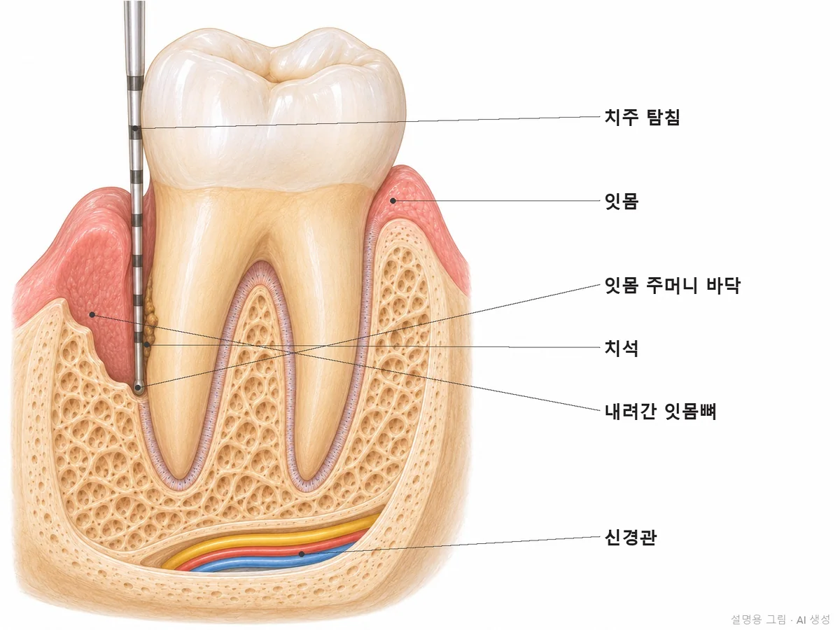
Gum exam
Gum exam. With a thin marked instrument, we measure the gum pocket depth of each tooth in millimeters and record bleeding sites and mobility.
STEP 01
We measure the gums first
Even for an implant consultation, we first check gum depth, mobility, and bleeding sites for all remaining teeth. We also look at all teeth and the gum bone with a digital panoramic X-ray.
Why we do thisIf implants are placed in a mouth with gum disease, the same inflammation easily develops around the implants. For implants to last, the surrounding gums must be healthy first, so we do gum treatment first if needed.
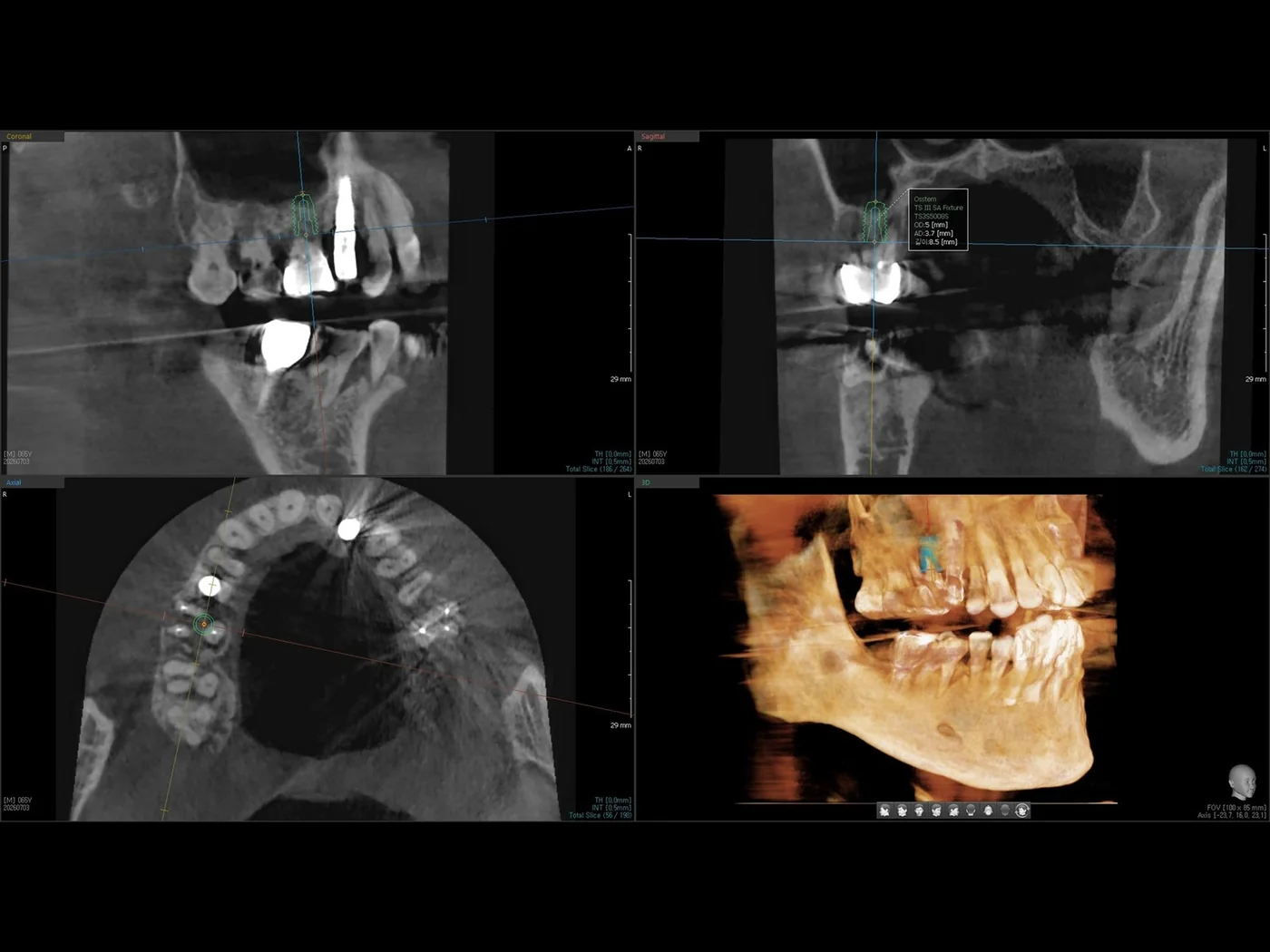
CT measurement
3D CT cross-section measurement. We measure in millimeters the bone height and width at the site and the remaining distance to the lower jaw nerve or the maxillary sinus.
STEP 02
We measure bone to the millimeter with 3D CT
With Vatech 3D CT, we measure the bone height and width at the site, bone density, and the distance to the lower jaw nerve canal and the maxillary sinus above. If bone is lacking, we show you on screen at this stage whether a bone graft is needed and how much.
Why we do thisA panoramic X-ray is a flat image, so it cannot show bone width, and distances look slightly different from reality. Implants must be placed at a safe distance from the lower jaw nerve, so measuring directly on 3D cross-sections is more accurate.
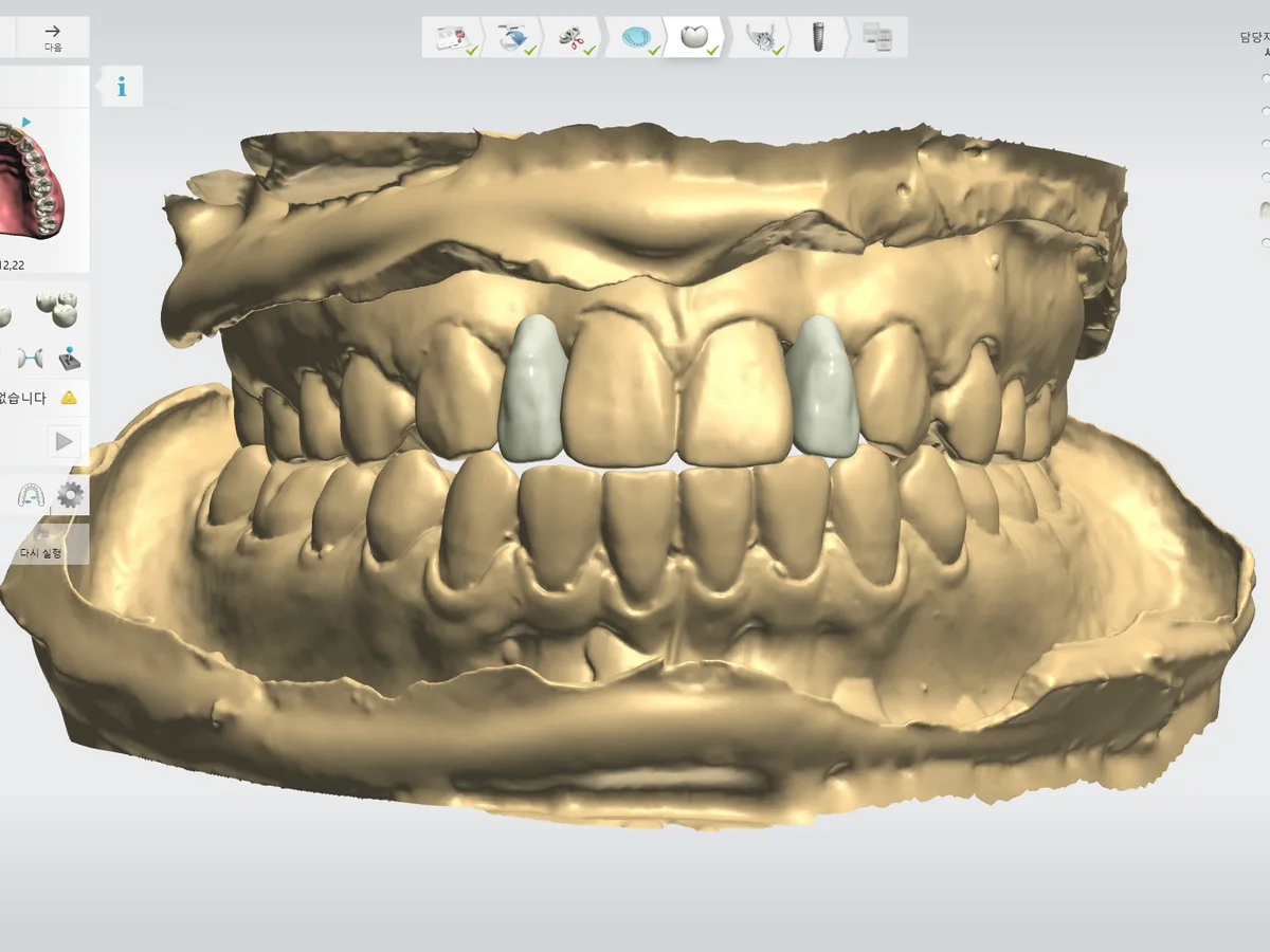
Tooth shape
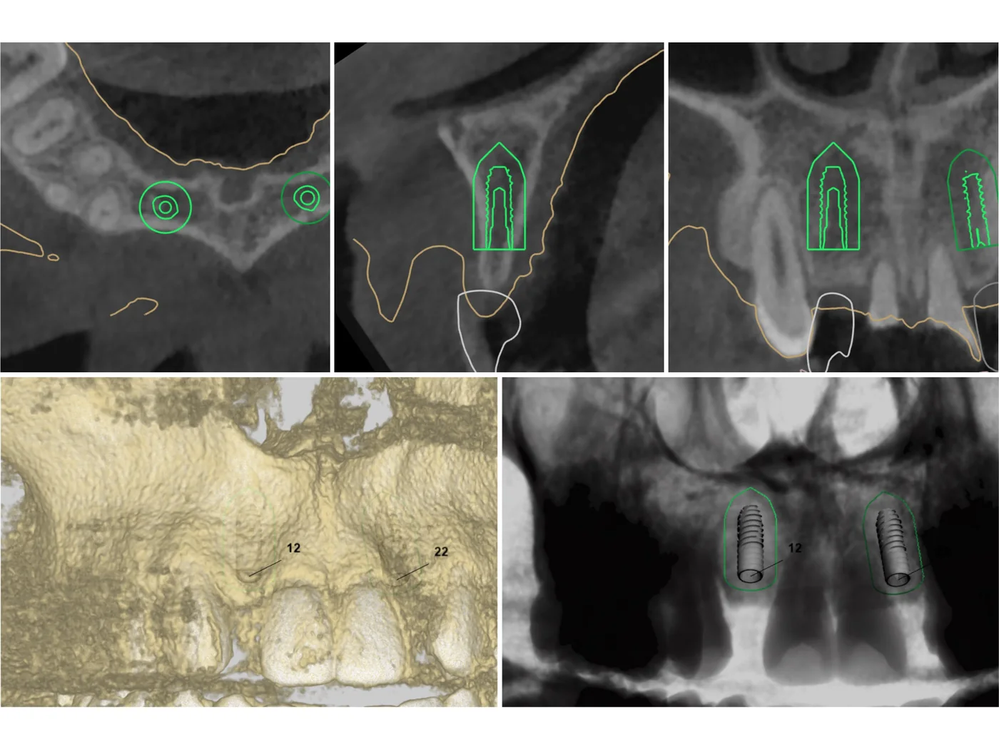
Position and angle
Guide
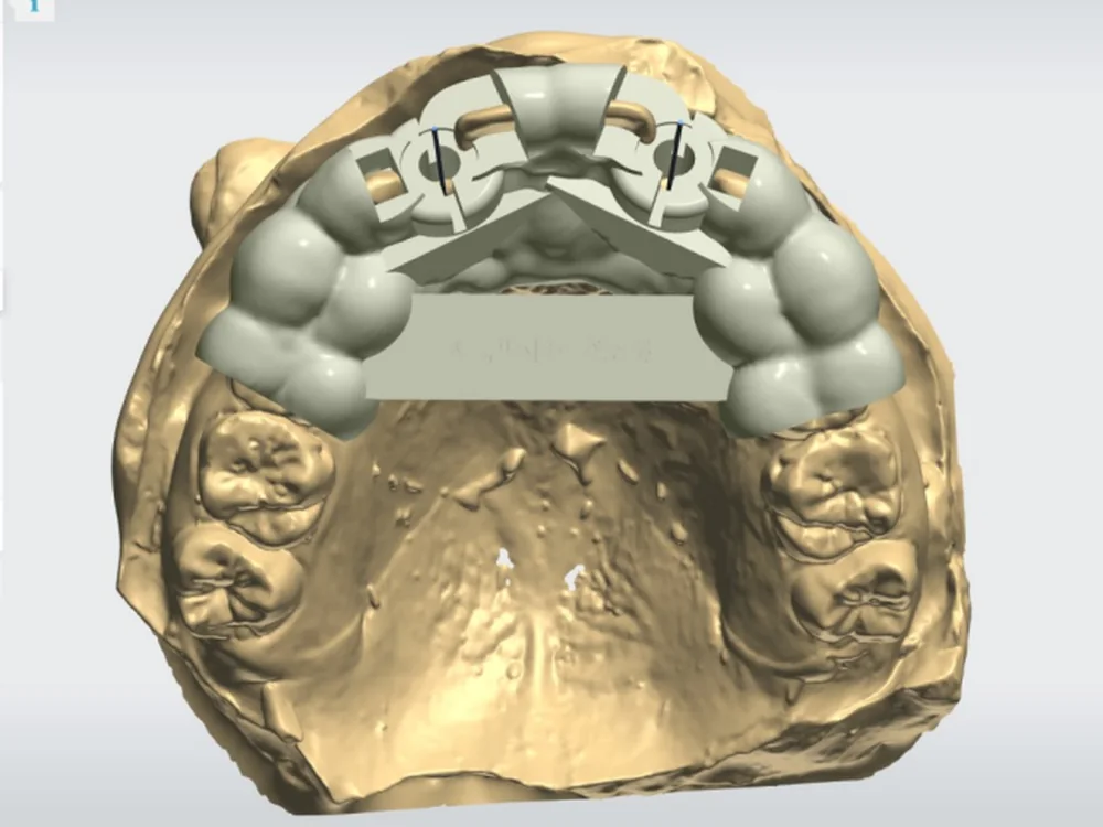
Design and navigation guide. We first draw the shape of the tooth to be placed, overlay the CT and intraoral scan to set the position, angle, and depth that will support that tooth, and then design a template with holes so the implant can be placed exactly as planned. All three screens are real design screens for the same patient.
STEP 03
We decide the shape of the tooth on top first, then work backward to the root position
With the iTero intraoral scanner, we save your teeth in 3D without traditional impressions. We first draw where the final tooth will sit, then place the implant at the position and angle that best supports that tooth. The navigation guide is made from this plan. We use a guide especially when several implants are needed, or when the bone is shaped unusually and there is no other place to put the implant.
Why we do thisIf an implant is placed simply wherever there is bone, the tooth on top may end up tilted or shaped in a way that is hard to clean. Biting force must travel straight down the implant post to reduce screw loosening and bone loss.
Placement
Right after placement
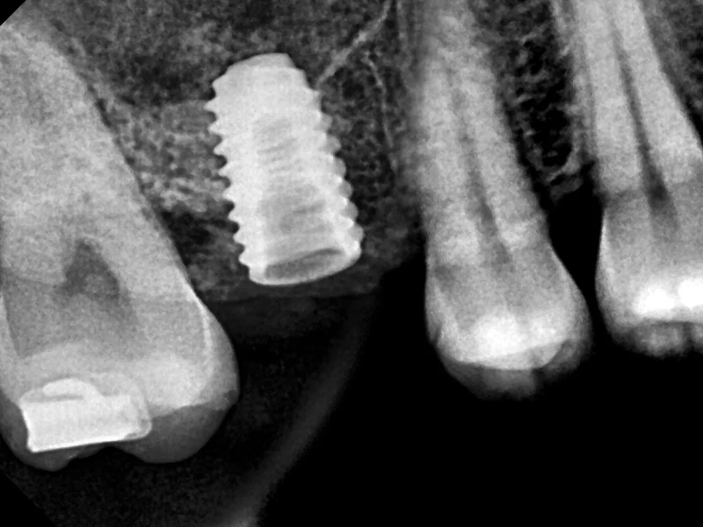
Placement and X-ray right after. After local anesthesia, we fit the guide and place the implant, then take an X-ray to confirm it went into the planned position.
STEP 04
We place implants under local anesthesia and check with an X-ray right away
Under local anesthesia, we place the implant at the planned site. One implant usually takes about an hour, longer with a bone graft. Afterward we take an X-ray to check the position, disinfect the site the next day, and remove stitches 1–2 weeks later.
Why we do thisHow firmly the implant grips the bone at the moment of placement (primary stability) affects the waiting period and whether a temporary tooth can be placed right away. So we record the stability at placement and use it for the next steps.
Waiting period
Bone-bonding period. While bone bonds to the implant surface, the gums heal, and we check the bone with X-rays at check-ups.
STEP 05
We wait until bone bonds to the implant
Bone usually takes about 2–3 months to bond to the implant surface in the lower jaw and about 3–4 months in the softer upper jaw, and longer if a bone graft was done. For visible sites such as front teeth, we provide a temporary tooth during this period.
Why we do thisIf strong biting force is applied before bone has fully bonded, the implant can loosen and fail. Rather than shortening the period, we choose to confirm bonding before moving on.
Post connection
Final crown
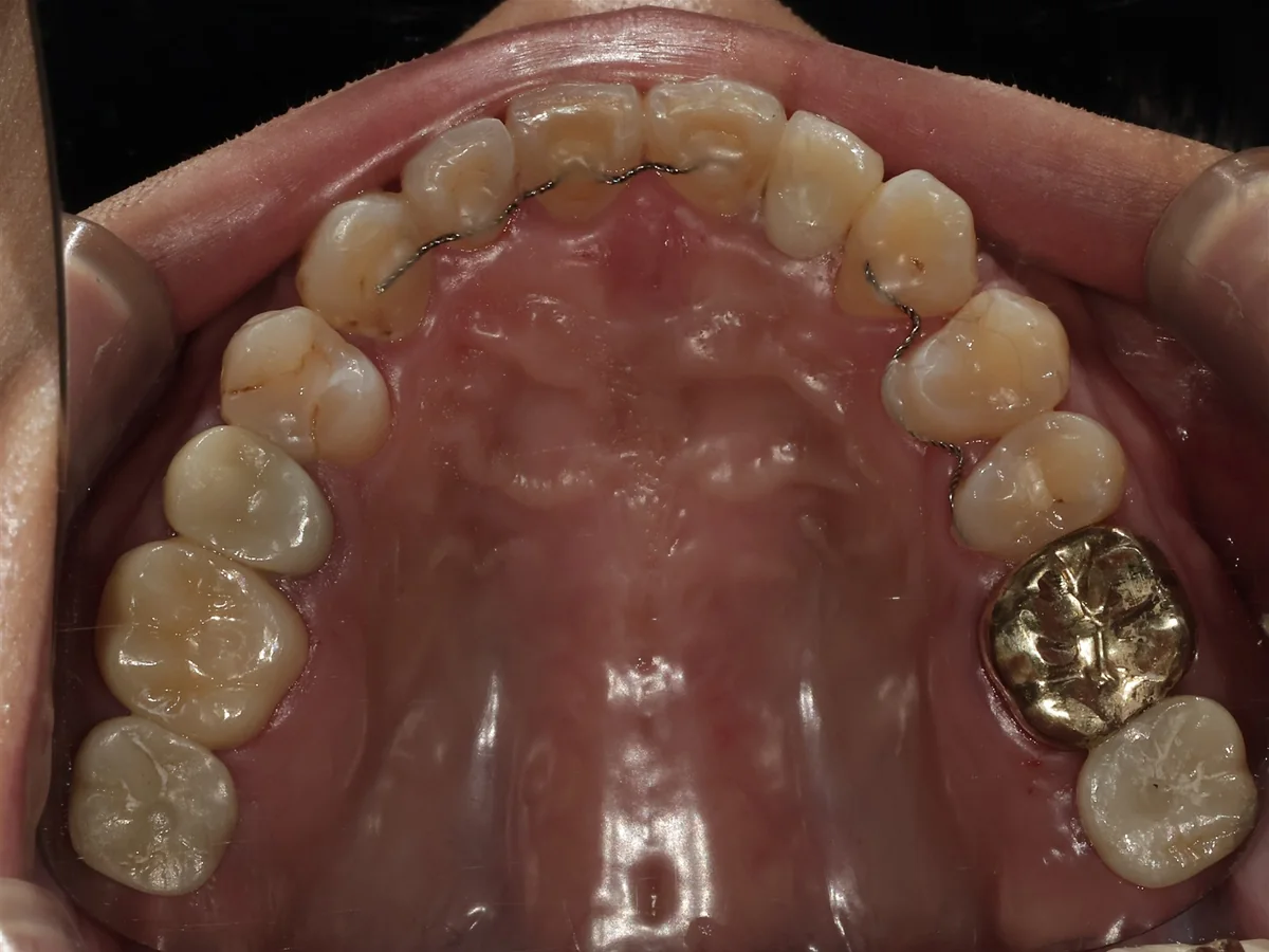
Crown. We connect a post to the implant, make the tooth from an intraoral scan, and fit it with the biting height adjusted. In the photo, the upper left second molar (#27) is the implant.
STEP 06
The same dentist places the crown and adjusts the biting height
Once bone bonding is confirmed, we connect the post (abutment) and make the tooth for the top from an intraoral scan. After fitting, we check the biting height, contact with neighboring teeth, and whether an interdental brush fits through.
Why we do thisImplants do not have the ligament that absorbs shock like natural teeth. To last, they should be set to touch slightly less than natural teeth, with a path for cleaning tools.
Right after the crowns · Mar 2025
15 months later · Jun 2026
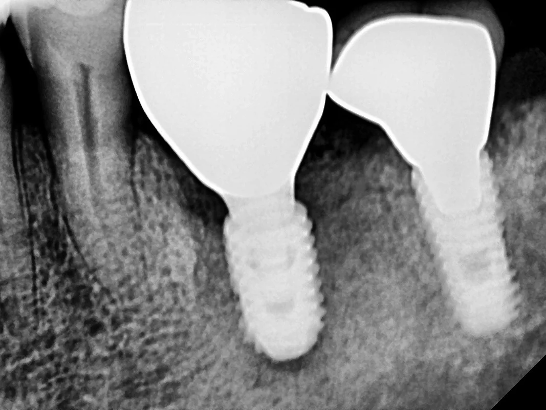
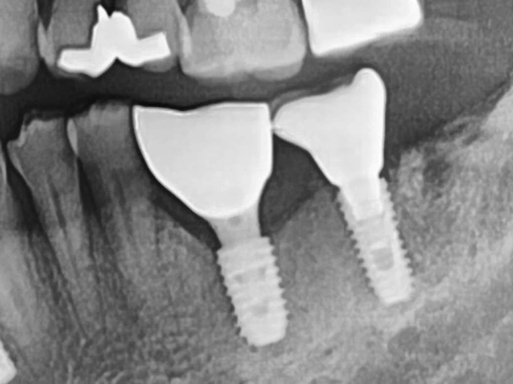
Regular check-up. We place the X-ray taken right after the crowns side by side with the panoramic X-ray 15 months later to compare whether the bone height around the implants has been maintained.
STEP 07
We check on a set schedule
Check-ups are at 1, 3, and 6 months after the crown, then every 6 months. People who have had gum disease, smoke, have diabetes, or have several implants are seen every 3 months. At check-ups, we check gum depth around the implant and bleeding or pus, and if needed compare bone height with the first X-ray.
Why we do thisImplants do not get cavities, but the gums and bone around them can become inflamed (peri-implantitis), and because it does not hurt early on, it is hard to notice yourself. That is why we think how the implant is followed for years afterward matters more than the day it is placed.
* Clinical photos were published with the patients' consent. Side effects such as temporary pain or discomfort may occur depending on the person.
For 2–3 days after surgery you may have swelling and soreness. Smoking makes it harder for bone to bond, so we recommend quitting before and after surgery.
06°
Options for a missing tooth compared
Saving the tooth
Dental Implants
Bridge
Partial denture
Good for
Teeth that can be kept with gum treatment or root canal treatment
Teeth that are missing or cannot be saved, at sites with bone or where bone can be built
When the teeth on both sides of the gap already have large restorations and need crowns
People with several missing teeth for whom surgery is difficult
Neighboring teeth
Not touched
Not touched
Teeth on both sides are shaped and connected
Supported by clasps hooked onto remaining teeth
Surgery
Gum surgery if needed
Needed (bone graft added if bone is lacking)
None
None
Duration
Depends on the extent of treatment
Usually a few months (longer with a bone graft)
Usually a few weeks
Usually a few weeks
Limits
If much bone has been lost, the tooth may not last long
Peri-implantitis care needed
Shaped neighboring teeth may later need cavity or root canal treatment
Must be removed for cleaning; weaker biting force
Korean National Health Insurance (NHI)
Covers most gum treatment and root canal treatment
For these people, another treatment comes first, or treatment is not needed
Good for you if
You have a gap where a tooth is missing
You have a tooth that cannot be saved because the root is cracked or decay has reached below the gums
A bridge would require shaping healthy neighboring teeth
Other treatment comes first
Gum inflammation: we do gum treatment first.
Lack of bone: we build the site first with a bone graft.
Taking medication for diabetes, osteoporosis, or similar: we check how well it is controlled and the type of medication, and proceed after getting your physician's opinion if needed.
You may not need it now
A tooth that can be saved: we show you the gum depth, mobility, and bone height and explain the option of keeping the tooth first.
08°
Is it possible to place an implant on the day of extraction?
We consider it only when there is little inflammation at the extraction site and enough bone remains to hold the implant firmly. We assess this in advance with CT, and if the conditions are not met, we place the implant after the extraction site heals.
Placing the implant on the day of extraction (immediate placement) and also placing a temporary tooth that day (immediate temporary tooth) require different conditions.
At sites with pus or where the bone wall has largely collapsed, it is safer to wait before placing the implant.
Front teeth can be kept out of biting, and a visible gap is uncomfortable, so for front teeth we usually place the implant and a temporary tooth on the day of extraction. For molars, it depends on how much bone remains.
09°
How we follow up after placement
Implants do not get cavities, but the gums and bone around them can develop disease just like natural teeth.
1
The day after surgery
We disinfect the site and check for bleeding and swelling.
2
1–2 weeks later
Stitches are removed.
3
1 month after the crown is fitted
We check the biting height (occlusion) and how clean the area is.
4
Then at 3 and 6 months
We check again at 3 and 6 months after the crown.
5
Every 6 months after that
Regular check-ups continue.
6
Every 3 months for higher-risk patients
People who have had gum disease (periodontitis), smoke, have diabetes, or have several implants are checked every 3 months.
What we do at check-ups: measure gum depth around the implant, check for gum bleeding and pus, compare bone height with X-rays if needed, professional cleaning, adjust the biting height, and review your home cleaning tools. At home, we recommend a soft toothbrush and interdental brushes, plus a Waterpik if needed.
10°
Equipment and products we use
Vatech 3D CT: measures bone height and thickness and the distance to nerves and the maxillary sinus in millimeters.
Digital panoramic X-ray: shows all teeth and the gum bone in one image.
iTero intraoral scanner: saves your teeth in 3D without traditional impressions, for guide and crown design.
Navigation implant guide: a surgical template that helps place implants at the position, angle, and depth set from CT and intraoral scans.
We use Osstem implants.
Sterilization: used surgical instruments are cleaned, packed one set at a time, and sterilized in a high-pressure steam sterilizer (autoclave).
11°
Self-check: do you need care now?
If even one applies, we will check your current condition with a gum exam and CT. If a gap is left long after extraction, the bone there can shrink and neighboring teeth can tilt.
12°
Side effects and limits
Please read
· For 2–3 days after surgery, you may have swelling, soreness, and bruising.
· Rarely, an implant does not bond to the bone and must be removed and placed again. Smoking and uncontrolled diabetes raise this risk.
· The gums and bone around an implant can become inflamed (peri-implantitis). If not managed, bone can be lost and the implant may be lost, so regular check-ups are needed.
· At lower molar sites, proximity to the nerve can rarely cause reduced sensation in the lip or chin. We therefore measure the distance with CT and keep a safe margin.
· The crown can break or the screw can loosen. In that case, we retighten it or make a new crown.
· Food easily gets caught between an implant and the neighboring tooth, so you need to use an interdental brush.
· If bone is lacking, a bone graft is needed and the whole treatment takes longer.
FAQ
FAQ
Will you recommend an implant even if my tooth can be saved?
Before any extraction, Ceramic Dental Clinic measures gum depth, mobility, and bone height, shows them to you on screen, and explains both the option of saving the tooth and the option of an implant. For teeth loose from gum disease, we often measure again after gum treatment and then decide.
Does implant surgery hurt?
Implants are placed under local anesthesia, so pain during surgery is usually not severe. For 2–3 days after surgery you may have swelling and soreness, so we prescribe anti-inflammatory painkillers, antibiotics, and digestive medicine.
After the implant is placed, bone usually takes about 2–3 months to bond in the lower jaw and about 3–4 months in the upper jaw, and then the crown is placed. With a bone graft, it takes a few more months.
If there is little inflammation at the extraction site and enough bone remains to hold the implant firmly, we consider immediate placement on the day of extraction. We assess this in advance with CT, and if the conditions are not met, we place the implant after the extraction site heals.
What implants does Ceramic Dental Clinic use?
Ceramic Dental Clinic uses Osstem implants. The product and price are listed together on the treatment cost guide page.
Can I get implants if I have diabetes or high blood pressure?
If your diabetes and high blood pressure are well controlled, implants are often possible. High blood sugar makes it harder for bone to bond and raises the risk of infection, so we check how well it is controlled first and get your physician's opinion if needed.
I take osteoporosis medication. Can I get implants?
Some osteoporosis medications can slow healing of wounds in the jawbone, so please be sure to tell us the name of the medication and how long you have taken it. Depending on the type and duration, we decide on timing after consulting the doctor who prescribed it.
Can implants get gum disease too?
The gums and bone around an implant can also become inflamed (peri-implantitis), and because it does not hurt early on, it is easy to miss. That is why we check at 1, 3, and 6 months after the crown, then every 6 months (every 3 months if risk is higher).
Do you also check implants placed at another clinic?
Yes. We check implants placed at other clinics too, and treat peri-implantitis if present. We first check the condition with X-rays and a gum exam. Compatible parts differ by product, so please bring records from your previous clinic if you have them.
Evidence and references
FDI World Dental Federation consensus (2019): recommends implant check-up intervals of 6 months or less
Academy of Osseointegration and American Academy of Periodontology consensus (AO/AAP, published 2025): risk factors for peri-implant disease (history of periodontitis, smoking, uncontrolled diabetes, etc.) and the need for ongoing maintenance. doi:10.1002/JPER.25-0270
Questions from patients who travel to Seoul for treatment.
How much does an implant cost?
OSSTEM or Dentium implants are KRW 1,200,000 per implant, and implants with a zirconia crown are KRW 1,500,000. If bone is lacking, a bone graft is KRW 200,000 to 1,000,000 per site, depending on your jawbone. International and Korean patients pay the same fees. See our fee schedule.
How many visits do I need, and how long should I stay in Korea?
Implants need time for the bone and the implant to bond firmly. So you either visit Korea more than once or plan a longer stay. The schedule depends on your bone and whether a bone graft is needed, and we explain it after a CT scan.
Can I have my consultation in English?
Yes. The doctor in charge of each field consults with you directly in English. For implants and gum care, this is Dr. Ilseop Ahn, a board-certified periodontist. Before you travel, you can send photos of your teeth on WhatsApp or by email. An accurate diagnosis is made after we examine you in person.
What happens after I go home?
Implants do not decay, but the gums around them can become inflamed. If you notice swelling, bleeding or movement, it needs to be checked. Send us a photo on WhatsApp if anything worries you. Regular check-ups and scaling help keep the area healthy.
Results, the number of visits and schedules vary from person to person. More questions: Visiting from abroad.


 Before · Jun 2020
 Day of gum treatment · Oct 2020
 3 years 3 months later · Jan 2024
 Before · Aug 2023
 13 days after placement · Apr 2024
 5 and a half months later · Oct 2024
 Before · Jul 2020
 Check-up · Jun 2022
 Gum exam
 CT measurement
 Tooth shape
 Position and angle
 Guide
 Placement
 Right after placement
 Waiting period
 Post connection
 Final crown
 Right after the crowns · Mar 2025
 15 months later · Jun 2026