Last reviewed October 29, 2026 · Dr. Ilseop Ahn (board-certified periodontist, specialist in integrated dentistry)
At a glance
Good for
Your jawbone is thin or low, so there is not enough room for an implant
Treatment flow
Measure the shortfall on a 3D CT → Bone graft (same day as placement if needed) → Implant placement → Final crown
Duration and limits
If the bone is built first, the implant is usually placed 4–6 months later, so treatment takes longer
Details
Even if your jawbone is thin or low, implants are often possible once bone grafting creates enough room. If the bone is not wide enough, we do guided bone regeneration, adding bone beside the implant as it is placed. If the bone at an upper molar site is too low below the air space above it (maxillary sinus), we do a sinus lift, raising the floor of the sinus. If enough bone remains, grafting and implant placement are done on the same day; if too little remains, we build the bone first and place the implant usually about 4–6 months later. At Ceramic Dental Clinic, a board-certified periodontist who specialized in jawbone regeneration measures the shortfall in mm on a 3D CT and shows it to you, and the same doctor handles everything from grafting to placement and the final crown.
01°
Depending on how much bone was missing, the method and order differed
Even "bone grafting" means different work depending on where and how much bone is missing. We show three cases with real X-rays and illustrations.
CASE01
Upper molar with very little bone left below the sinus
Sinus lift (lateral approach) to build bone height first, then implant placement once the bone had filled in
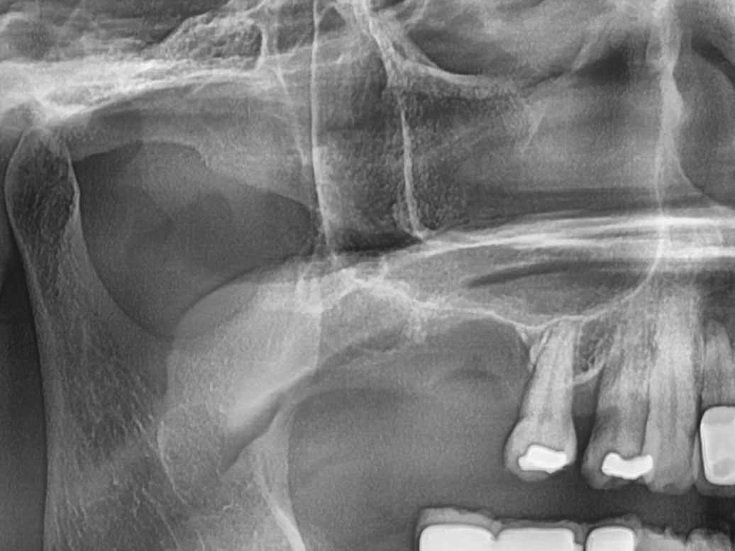
Before · Jun 2024
X-ray before treatment (panoramic). Little bone remained between the upper right molar site and the sinus floor, so it would have been hard to hold an implant firmly as it was.
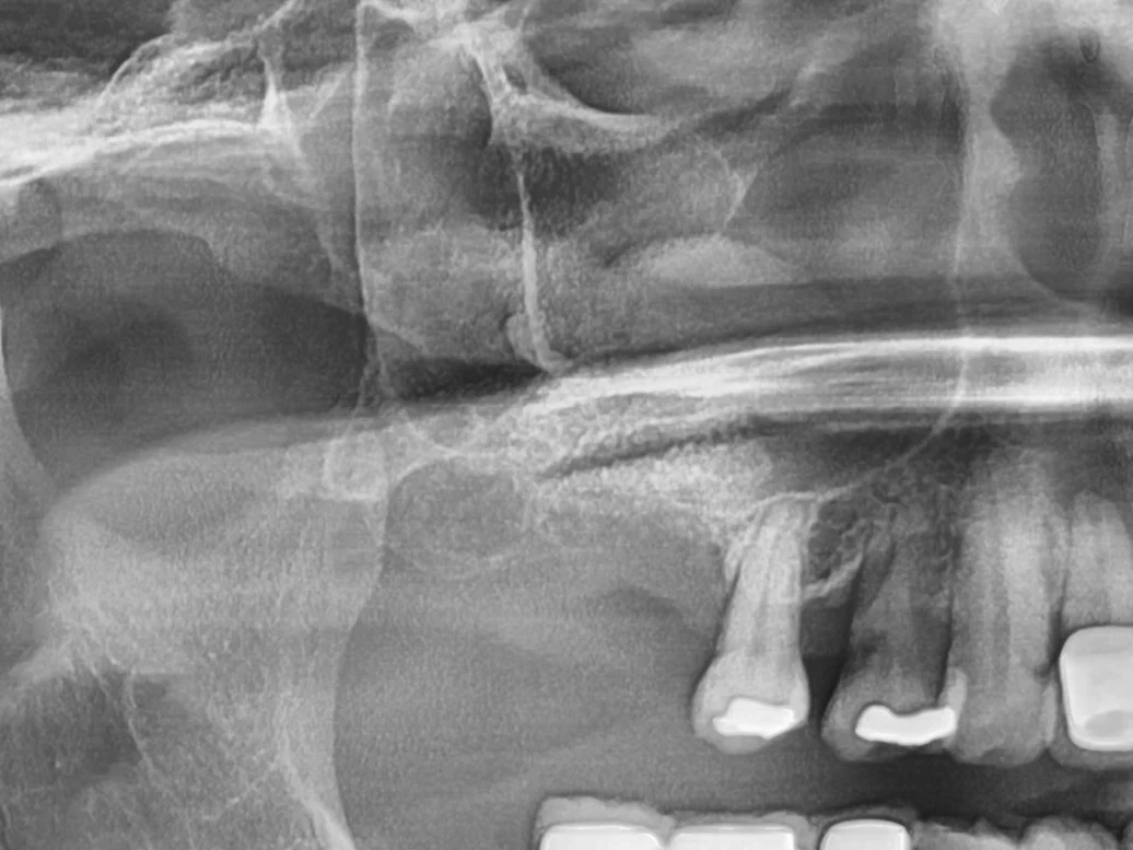
1 week after grafting · Sep 2024
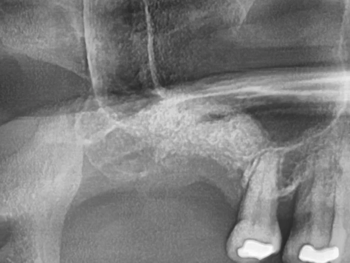
X-ray 1 week after grafting. A small window was opened on the side of the gum, the sinus floor was raised, and graft material was packed in. A white mass of graft material sits above the sinus floor.
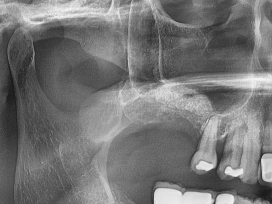
4 months after grafting · Jan 2025
X-ray 4 months after grafting. The raised height is being maintained. It takes about 5–6 months for graft material to turn into bone, so we decided to check once more with a CT before placing the implant.
Condition
Missing upper molar, about 2mm of remaining bone height
Chosen
Implant placed 6 months after a sinus lift through a side window
Not done
Same-day placement (too little bone remained for good initial stability)
Total duration
9 months
Note
Even after the implant is placed, the bone at a grafted site must be monitored with regular X-rays.
CASE02
Upper molar with slightly too little bone
Sinus floor raised slightly through the implant hole, with same-day placement
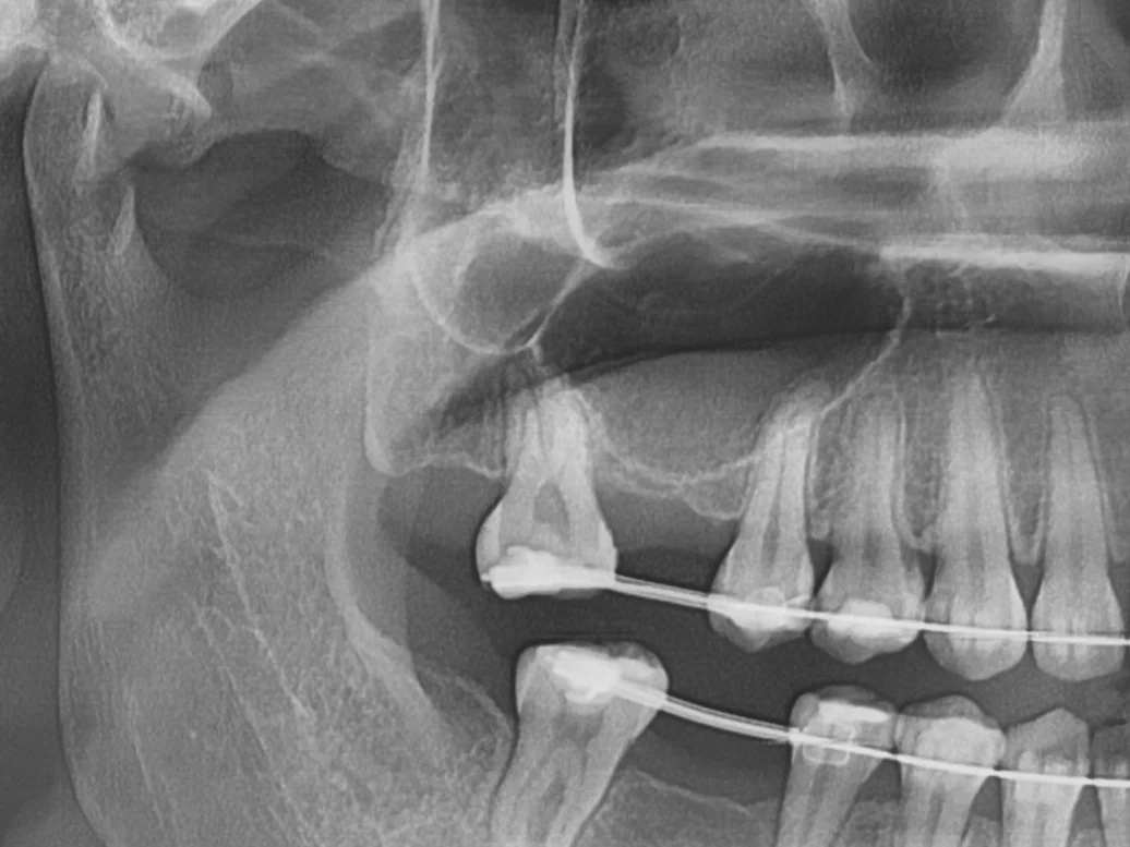
Before · Aug 2023
X-ray before treatment (panoramic). The bone at the upper right first molar site was low because of the sinus and slightly shorter than the implant length. There was still enough to hold the implant when placed.
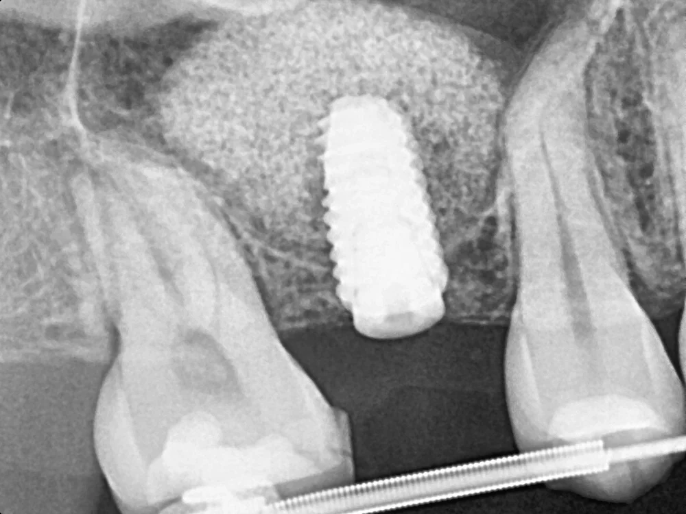
13 days after placement · Apr 2024
X-ray 13 days after placement. The sinus floor was raised slightly through the implant hole and graft material was added, creating a rounded dome shape above the implant tip.
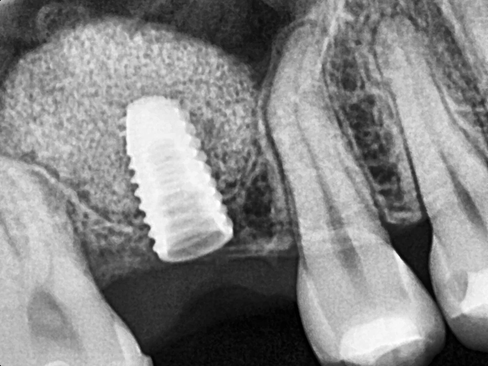
5 and a half months later · Oct 2024
X-ray 5 and a half months later. We confirmed that the dome-shaped graft area remained in place and took impressions for the crown.
Condition
Upper molar, about 3mm of remaining bone height
Chosen
Sinus floor raised through the implant site hole, with same-day placement
Not done
Major surgery with a side window (only a small lift was needed)
Duration
About 6 months from placement to crown impressions
CASE03
Thin bone that nearly left the side of the implant exposed
Guided bone regeneration during placement: adding bone to the thin side and covering it with a membrane
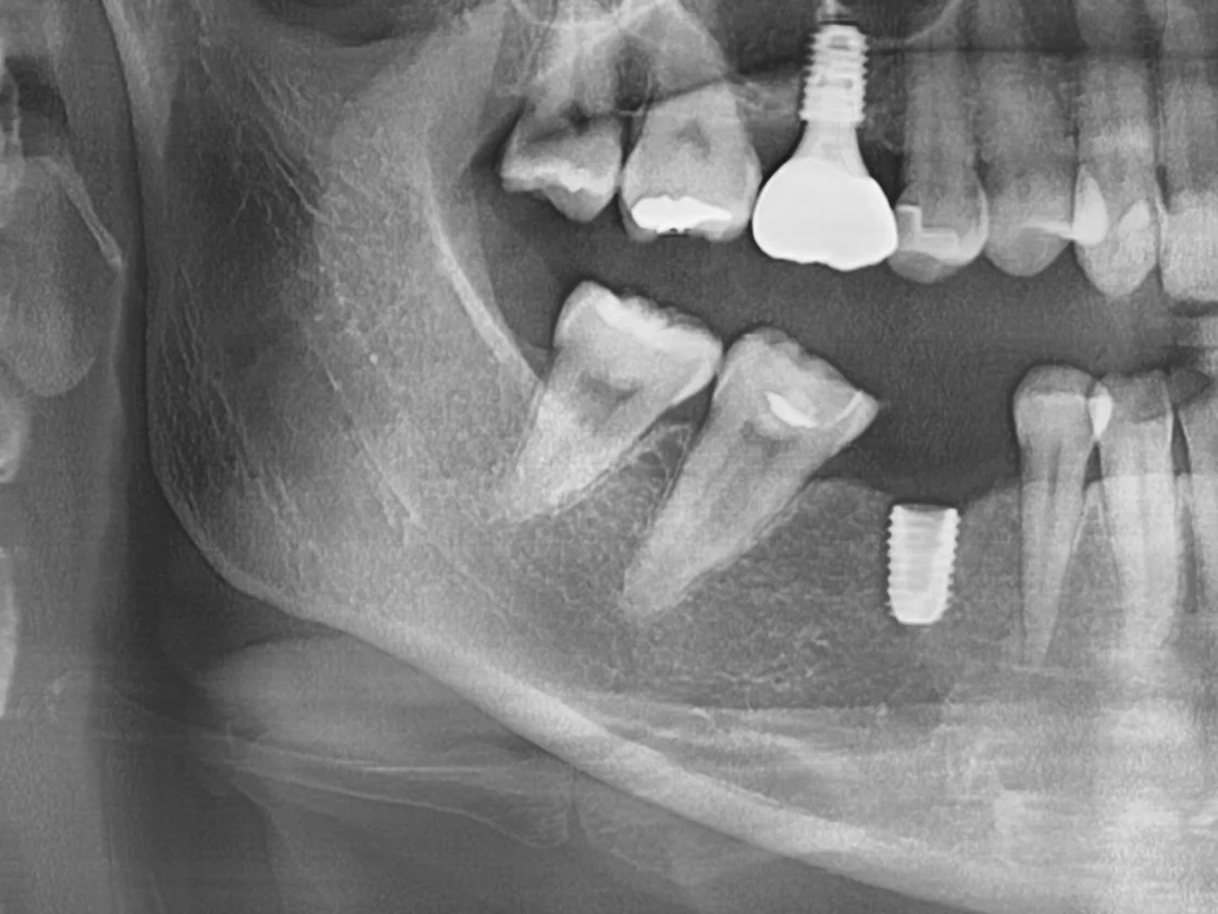
Day of surgery · Jul 2024
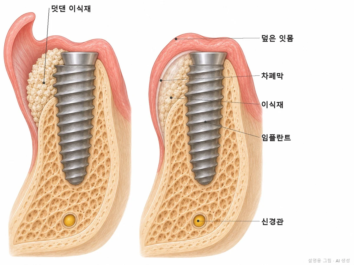
X-ray on the day of surgery (panoramic), right after an implant was placed at the lower right first molar site. On an X-ray, the cheek side and tongue side overlap in one image, so bone "width," the problem in this case, does not show. That is why the surgery is explained with the two illustrations beside it.
Cross-section
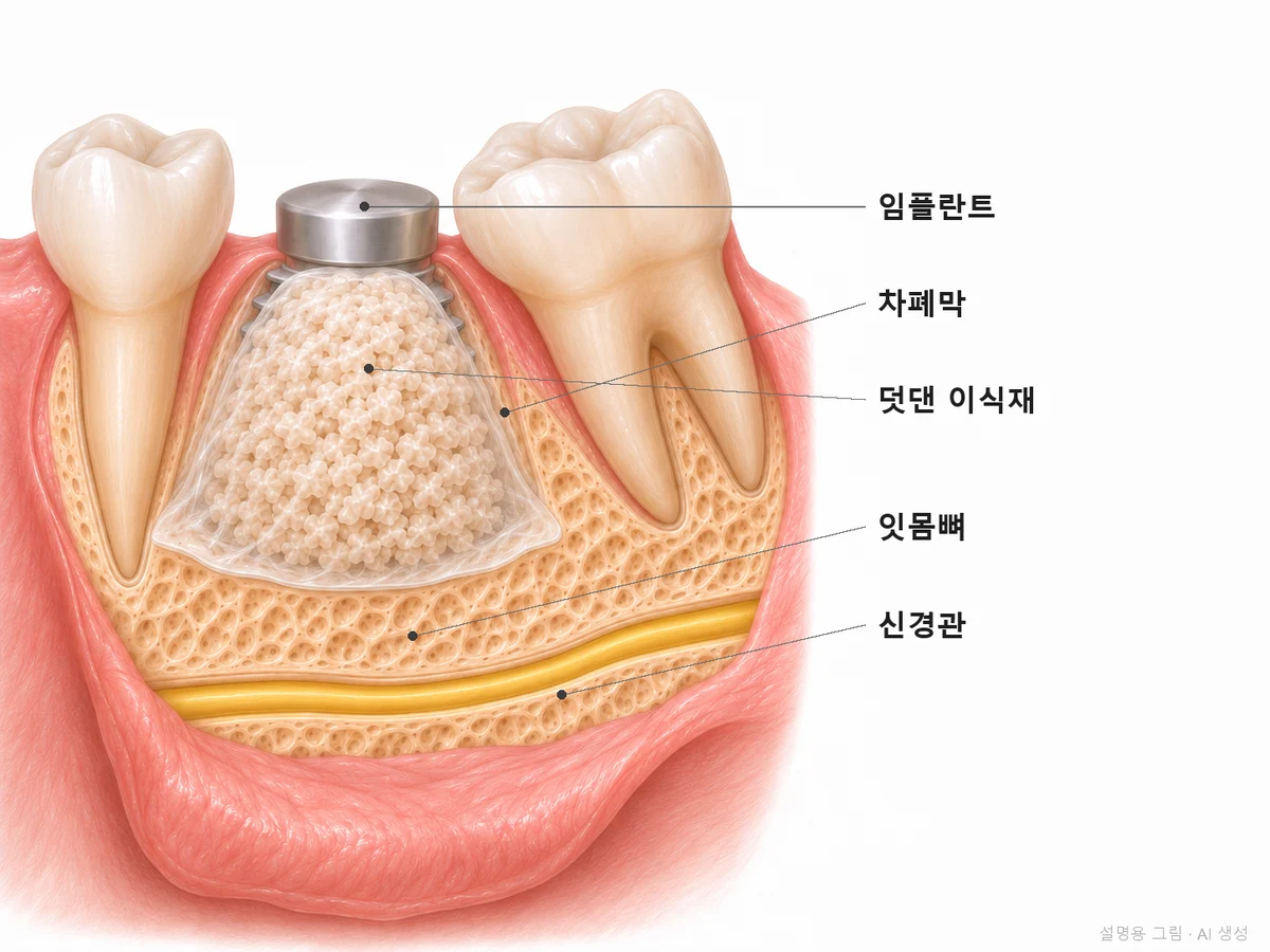
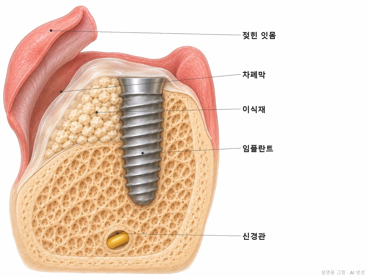
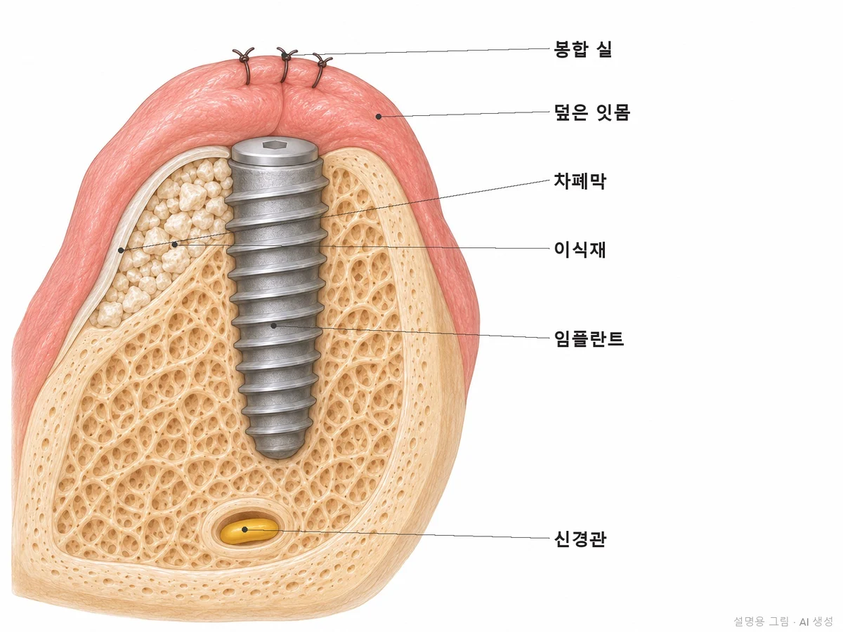
Cross-section of the same site cut from the front. Where the bone was thin and the implant threads on the cheek side were exposed outside the bone, graft material was added (left) and covered with a barrier membrane and gum (right).
View from the cheek side
View from the cheek side. Graft material was wrapped over the exposed threads and covered with a barrier membrane so that gum tissue would not grow into the space where bone should form.
2 months later · Sep 2024
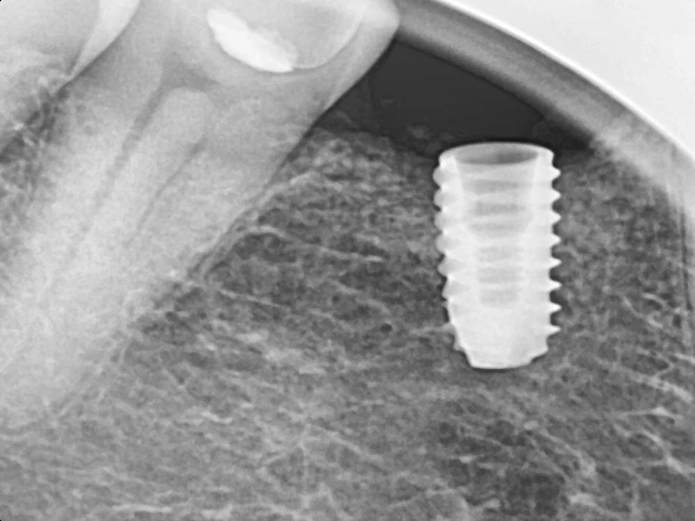
X-ray 2 months later. The implant had settled, so impressions for the crown were taken that day, and the crown was placed in October 2024. The width of the added bone can only be measured on a CT, so it is not compared in this photo.
Condition
Thin bone, about 4mm wide
Chosen
Implant placement and guided bone regeneration on the same day
Not done
Separate surgery to build bone first (height was enough)
Duration
About 5 months from surgery to crown
* Photos were taken under the same conditions and published with the patients' consent. Treatment results and side effects may vary from person to person.
02°
Why does jawbone become insufficient?
Depending on the cause, the direction (height or width) and amount of bone loss differ.
01Time has passed since a tooth was removed
The bone that supported a tooth shrinks once the tooth is gone. Width shrinks a lot especially in the first few months after extraction, and the longer the gap is left, the thinner the bone tends to become.
02A tooth was lost to gum disease
Gum disease destroys the bone that supports the teeth, so by the time a tooth is lost, the bone height has often already dropped a lot.
03Long-term denture use
If a denture presses on the gums for a long time, the bone underneath can gradually become lower and thinner.
04Upper molar sites
Above the upper molars is an air space called the maxillary sinus. When a molar is lost, the sinus can drop lower, leaving even less bone height.
03°
How implants with bone grafting work
Six steps from CT measurement to checkups. We explain what we look at in each step and why, with real clinical photos.
CT measurement
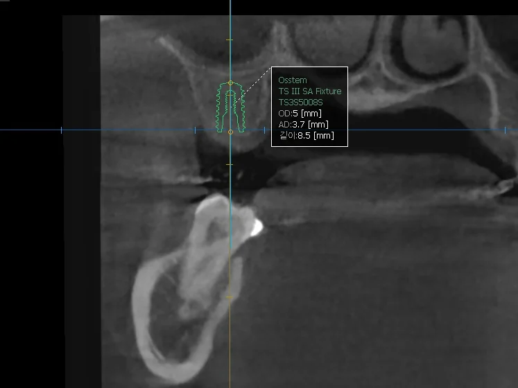
3D CT measurement. We place a virtual implant (5mm wide, 8.5mm long) at the planned site and check the remaining bone height and the distance to the sinus, so the shortfall is judged in numbers.
STEP 01
We measure the shortfall in numbers
We measure bone height and width at the planned site with a Vatech 3D CT. To surround a typical implant, a width of usually around 6mm and a height of about 8–10mm makes things easier. If there is less, we show you on screen in which direction and by how much it falls short.
Why we do this"You have no bone" alone does not tell you what needs to be done. The method and duration differ completely depending on whether height or width is lacking, and by how much.
Sinus check
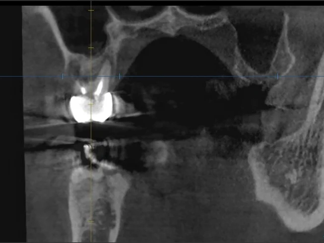
Sinus check. On a CT cross-section, we look at whether the membrane lining the sinus (dark air space) above the molar site is thickened and swollen, or filled with inflammation.
STEP 02
For upper molars, we check the sinus first
A sinus lift raises the thin membrane on the sinus floor and fills the space beneath it with graft material. If the membrane is thickened and swollen, or there is inflammation such as sinusitis, we first recommend seeing an ENT doctor and postpone the surgery.
Why we do thisOperating on an inflamed sinus can make inflammation worse after surgery or infect the graft material. Checking the membrane before surgery is the surest prevention.
Choosing the method
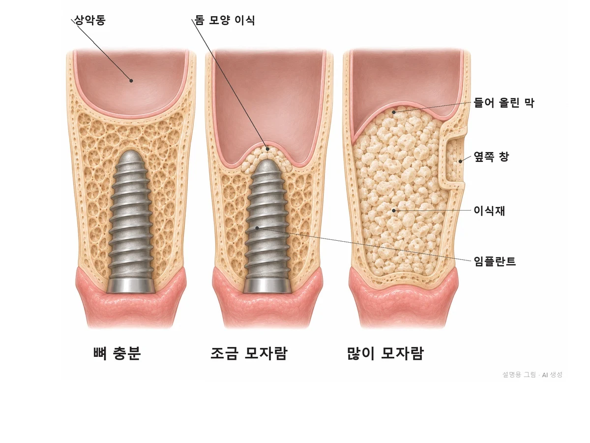
Choosing the method and order. If there is enough bone, the implant is placed as is. If bone is slightly short, the sinus floor is raised in a dome shape through the implant hole and the implant is placed the same day. If bone is very short, the floor is raised a lot through a side window to build bone first.
STEP 03
We decide whether to do it the same day or build bone first
If the remaining bone alone can hold the implant firmly, grafting and placement are done on the same day. At upper molars, if roughly 5mm or more of bone remains, the sinus floor is often raised through the implant hole and the implant placed at the same time. If less remains, a small side window is often opened to raise the sinus floor, and we wait for the bone to fill in before placing the implant. The threshold depends on your bone quality (how dense the bone is) and is decided with a CT.
Why we do thisDoing it on the same day reduces the number of surgeries and the overall time, but placing an implant with weak stability raises the risk that it will not bond to the bone. We look at the conditions for the implant to bond before the number of surgeries.
6 weeks after grafting · Oct 2024
Barrier membrane
Stitching
Graft surgery. The first photo is an X-ray 6 weeks after the sinus floor was raised through a side window and filled with graft material; a mass of graft material is visible above the sinus floor (same patient as Case 1). As in the next two illustrations, the graft material is covered with a membrane and the gum is stitched so that gum tissue does not grow into the space first.
STEP 04
We fill in graft material and cover it with a membrane
Under local anesthesia, we fill the deficient site with graft material. A barrier membrane is placed over the graft material to keep gum tissue out and protect the space where bone will grow. The site is disinfected the next day, and stitches are removed after 1–2 weeks.
Why we do thisGum tissue grows faster than bone. Without a membrane, gum fills the space where bone should form first, and less bone forms than intended.
Right after grafting · Mar 2026
6 months later · Sep 2026
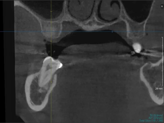
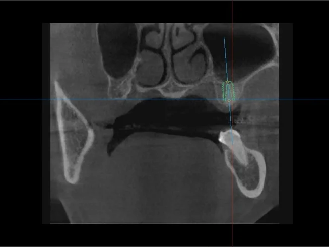
Time for bone to fill in. These are CTs of a site where bone was built first, right after grafting and 6 months later. On the 6-month CT, we confirmed that enough bone had filled in for an implant and placed it that same day. The two CTs were cut at slightly different angles, so the same heights cannot be compared directly.
STEP 05
We wait for the bone to fill in and place the implant after checking with a CT
When bone is built first, we usually wait about 4–6 months, check with a CT and then place the implant. Even with same-day placement, we wait a few months longer than for a standard implant before placing the crown. The time depends on the bone condition.
Why we do thisHow fast graft material turns into real bone differs by person and by site. We do not place the implant just because the date has come; we check with a CT before moving on.
Day the crown was placed · Oct 2024
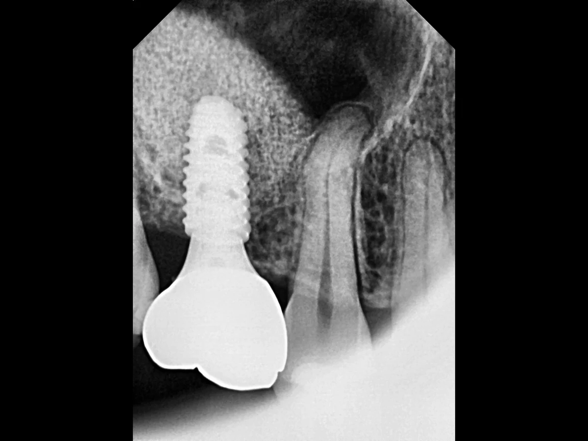
Crown and checkups. This X-ray was taken on the day the crown was placed on the implant of the Case 2 patient. The dome-shaped graft area above the implant tip remains in place, and at future checkups we compare with this photo to see whether the bone height is maintained.
STEP 06
The same doctor places the crown and does the checkups
Once we confirm the bone has bonded, we place the crown and check at 1 month, 3 months and 6 months after the crown, then every 6 months. At grafted sites, we compare the bone height on X-rays with the starting point.
Why we do thisGrafted bone can also shrink if inflammation develops. To keep the bone that was built for a long time, care after placement is as important as the surgery.
* Clinical photos were published with the patients' consent. Side effects such as temporary pain or discomfort may occur depending on the person.
04°
What types of bone grafting are there?
01Guided bone regeneration (GBR)
When bone width or height is slightly short, graft material is added and covered with a membrane to build bone. This is often done at the same time as implant placement.
02Sinus lift (crestal approach)
When an upper molar site is slightly short of bone, the sinus floor is raised a little through the hole drilled for the implant and graft material is added. The implant is usually placed the same day.
03Sinus lift (lateral approach)
When an upper molar site is very short of bone, a small window is opened on the side of the gum, the sinus floor is raised a lot, and graft material is packed in. The implant is often placed after the bone fills in.
04Socket preservation
When a tooth is removed, graft material is placed in the socket to reduce bone shrinkage. We consider this when an implant cannot be placed right away.
05°
Comparing methods
Grafting and placement on the same day
Grafting first, then placement
Short implant
Good for
When the remaining bone alone can hold the implant
When too little bone remains to hold the implant
When height is slightly short but width is enough and you want to avoid grafting
Number of surgeries
Once
Twice (grafting, placement)
Once
Total duration
A few months longer than a standard implant
Usually placement 4–6 months after grafting, then a few more months to the final crown
Similar to a standard implant
Limits
Not possible if stability is weak
Takes the longest
Cannot be used at every site and is less suitable when the tooth on top is long · Used occasionally at sites where conditions fit
Health insurance
Even with an NHI implant (for people enrolled in Korea's National Health Insurance), additional surgery such as bone grafting is not covered
For these people, another treatment comes first, or treatment is not needed
Good for you if
The CT shows your jawbone height or width is not enough
The bone below the sinus at an upper molar site is low
Another clinic told you implants are difficult because you have no bone
Other treatment comes first
You have gum inflammation: gum treatment comes first.
You have sinus inflammation (sinusitis): we first recommend ENT treatment.
You smoke: grafted bone does not bond well, so we first recommend quitting.
You may not need it now
The CT shows enough bone: we tell you that we graft only when the CT shows a shortfall, and do not graft if it is not needed.
07°
Precautions after surgery (especially after a sinus lift)
Do not blow your nose hard. Pressure inside the sinus can rise and damage the membrane.
Sneeze with your mouth open.
Avoid using straws and smoking. They change the pressure in your mouth and slow healing.
For 1–2 weeks after surgery, do not chew hard on the treated side.
Flying and diving: avoid flying and diving for at least 2 weeks after surgery. If you have travel plans, let us know before surgery.
Take prescribed medicine as directed.
08°
How we follow up after placement
Implants do not get cavities, but the gums and bone around them can become diseased like natural teeth. That is why we think how the implant is looked after in the years that follow matters more than the day it is placed.
1
Day after surgery
We disinfect the site and check bleeding and swelling.
2
1–2 weeks later
Stitches are removed.
3
1 month after the crown is placed
We check the bite height (occlusion) and how well it is being cleaned.
4
Then at 3 and 6 months
We check again 3 and 6 months after the crown.
5
Every 6 months after that
Regular checkups continue.
6
Every 3 months for higher-risk patients
If you have had gum disease (periodontitis), smoke, have diabetes or have several implants, we check every 3 months.
At checkups we: measure gum depth around the implant, check for gum bleeding and pus, compare bone height on X-rays if needed, do professional cleaning, adjust the bite height, and check the cleaning tools you use at home.
09°
Self-check
If any apply, we measure bone height and width on a CT and explain how many mm are missing.
10°
Side effects and limits
Please read
· Swelling and bruising may last from a few days to about 1 week after surgery, usually worst on day 2–3.
· Some graft material may be absorbed, so less bone than expected may form and an additional graft may be needed.
· If the grafted site becomes infected, the graft material may need to be removed and the graft redone.
· During a sinus lift, the membrane may tear (perforation). If small, it is repaired during surgery and the procedure continues; if large, the surgery is stopped and redone after it heals.
· For a few days after a sinus lift, you may have a slight nosebleed or a stuffy nose.
· Smoking and uncontrolled diabetes make it harder for bone to bond.
· Bone grafting adds a few months to the overall treatment time.
FAQ
FAQ
Do I have to get a bone graft for an implant?
We do bone grafting only when the CT shows that bone height or width is not enough. We show you on the CT screen why it is needed and how many mm are missing, and if it is not needed, we do not do it.
Even with too little bone, implants are often possible once a sinus lift or guided bone regeneration creates enough room. We take a new CT to check in which direction and how much bone is missing, and explain the options.
How much longer does an implant take with a bone graft?
If grafting and placement are done on the same day, it takes a few months longer than a standard implant. If the bone is built first, we usually wait 4–6 months before placing the implant, and then it takes a few more months to get to the final crown.
What materials are used for bone grafts?
Bone graft materials include processed human bone, processed animal bone and synthetic materials, and we choose based on the site and the amount needed.
Isn't a sinus lift risky?
A sinus lift carries a risk of tearing the sinus membrane or infection, so before surgery we check the membrane thickness and any inflammation on a CT. Depending on how much bone remains, we choose between raising the floor a little through the implant hole or opening a small window on the side to raise it a lot.
After a bone graft, swelling is usually worst on day 2–3 and mostly goes down within 1 week. If the area is large, bruising may also appear.
Is bone grafting covered by health insurance?
Even if you get an implant under Korea's National Health Insurance (NHI) for enrollees aged 65 or older, additional surgery such as bone grafting is not covered. Prices are on the treatment cost page.
Can a short implant help me avoid a bone graft?
If bone height is only slightly short and the width is enough, a short implant can sometimes avoid grafting. We also need to consider the length of the tooth to be placed on top and your biting force, so we decide based on the CT.
Evidence and references
FDI World Dental Federation consensus (2019): recommends implant checkup intervals of 6 months or less
Academy of Osseointegration and American Academy of Periodontology consensus (AO/AAP, published 2025): risk factors for peri-implant disease and the need for ongoing maintenance. doi:10.1002/JPER.25-0270
Questions from patients who travel to Seoul for treatment.
How much does an implant cost?
OSSTEM or Dentium implants are KRW 1,200,000 per implant, and implants with a zirconia crown are KRW 1,500,000. If bone is lacking, a bone graft is KRW 200,000 to 1,000,000 per site, depending on your jawbone. International and Korean patients pay the same fees. See our fee schedule.
How many visits do I need, and how long should I stay in Korea?
Implants need time for the bone and the implant to bond firmly. So you either visit Korea more than once or plan a longer stay. The schedule depends on your bone and whether a bone graft is needed, and we explain it after a CT scan.
Can I have my consultation in English?
Yes. The doctor in charge of each field consults with you directly in English. For implants and gum care, this is Dr. Ilseop Ahn, a board-certified periodontist. Before you travel, you can send photos of your teeth on WhatsApp or by email. An accurate diagnosis is made after we examine you in person.
What happens after I go home?
Implants do not decay, but the gums around them can become inflamed. If you notice swelling, bleeding or movement, it needs to be checked. Send us a photo on WhatsApp if anything worries you. Regular check-ups and scaling help keep the area healthy.
Results, the number of visits and schedules vary from person to person. More questions: Visiting from abroad.


 Before · Jun 2024
 1 week after grafting · Sep 2024
 4 months after grafting · Jan 2025
 Before · Aug 2023
 13 days after placement · Apr 2024
 5 and a half months later · Oct 2024
 Day of surgery · Jul 2024
 Cross-section
 View from the cheek side
 2 months later · Sep 2024
 CT measurement
 Sinus check
 Choosing the method
 6 weeks after grafting · Oct 2024
 Barrier membrane
 Stitching
 Right after grafting · Mar 2026
 6 months later · Sep 2026
 Day the crown was placed · Oct 2024